Saizjimenez Et Al 2011 Paleolithic Art in Peril Policy and Science Collide at Altamira Cave
Abstruse
The paintings from Tomba della Scimmia, in Tuscany, are representative of the heavy bacterial colonization experienced in most Etruscan necropolises. The tomb remained open until the belatedly seventy′s when it was airtight considering of severe deterioration of the walls, ceiling and paintings afterwards decades of visits. The deterioration is the upshot of environmental changes and impacts suffered since its discovery in 1846. Nosotros show scanning electron microscopy and molecular studies that reveal the extent and nature of the biodeterioration. Actinobacteria, mainly Nocardia and Pseudonocardia colonize and abound on the tomb walls and this process is linked to the availability of organic affair, phyllosilicates (e.g. dirt minerals) and iron oxides. Nocardia is plant metabolically active in the paintings. The data confirm the specialization of the genera Nocardia and Pseudonocardia in the colonization of subterranean niches.
Introduction
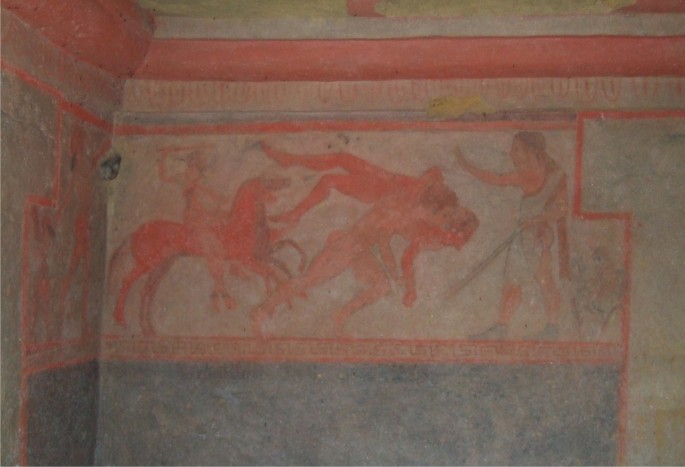
The Etruscan civilization was characterized by the construction of tombs decorated with impressive paintings1. The Tomba della Scimmia (Tomb of the Monkey) 480–470 BC, well-nigh Chiusi, Italy, discovered in 1846 by Alexander François, was named subsequently a monkey on a tree depicted in the archway sleeping room (Fig. 1). The tomb is composed of three rooms around an atrium and excavated viii m below the footing level on a lithological complex of Pliocene sands weakly cemented with intercalations of clay layers and pebble beds. The walls were decorated with paintings representing funeral games. The paintings were fabricated with hematite, Egyptian blueish and charcoal black, deposited on a previously prepared thin clay layertwo.

Painting in Tomba della Scimmia, Chiusi, Italy.
The current status of the tomb is the result of the accumulation of multiple microenvironmental changes and impacts suffered from the fourth dimension of its discovery. As many other Etruscan and Roman necropolises, this tomb is included in a tourism net that brings thousands of visitors per twelvemonth. Tombs receiving visits are exposed to microclimatic modifications due to the presence of visitors, which originated disturbances of the usual steady climate (i.e., switch on/off of the lights, openings of the door, ventilation, etc.).
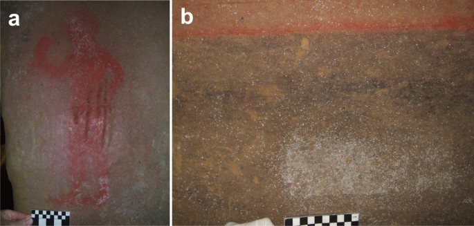
At the beginning of this century, the tomb ceiling presented a detached sand stratum, while cloth losses occurred between the sands and the underlying beds of pebbles, which was the point where the rock had a bottom cohesion. In addition, between 1971 and 1989 the wall surfaces were partly covered with whitish spots attributed to common salt efflorescences, merely they were after identified as bacterial colonizations. The walls were treated with Neodesogen (benzalkonium chloride) in a restoration carried out in 1993 and with 2% Preventol R80 (dodecyl dimethyl dichlorobenzyl ammonium chloride) in a further restoration in 2000. Currently the tomb is open to visitors for three days a week and 2 visits every 24-hour interval and the walls and paintings are heavily colonized past bacteria as shown in Effigy 2a,b.

Painting heavily colonized past actinobacteria.
(a) Sample SC3 was taken from the right leg. (b) Sample SC6 was taken from the whitish area.
The aim of this work is to know the reason for such a heavy bacterial colonization, exhibited by near Tuscanian tombs and the composition of the bacterial communities involved in the deterioration of the paintings of Tomba della Scimmia.
Results
Microscopic study
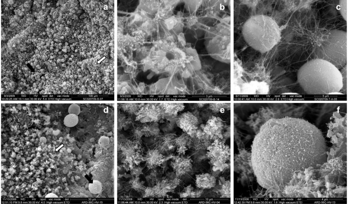
White colonizations were sampled from different rooms. In general, the colonizations are small, from less than 1 mm up to two–three mm diameter. They present an outline typically divers, circular to irregular and have petty relief. Unlike other subterranean environments3, the bacterial mats exercise not human activity as h2o condensation points. Environmental scanning electron microscopy (ESEM) showed that the colonizations were formed by a dumbo network of fine and large bacterial filaments with some extracellular polymeric substances (EPS) and a bed of mineral elements with morphology in rosette or "nest" and spherical elements (Fig. 3a–c). The morphology of the filaments is compatible with those of actinobacteriaiii and similar mineral structures were previously found in Altamira Cave, Spain4,v.

Environmental scanning electron micrographs of white colonizations from Tomba della Scimmia (a–c) and from Ardales Cave (d–f).
(a) Bed formed of CaCOiii nest-similar aggregates (white arrow) with dispersed spheroidal elements (black arrows) from Tomba della Scimmia. (b) Small patches of CaCO3 nest-like aggregates and filaments from Tomba della Scimmia. (c) Detail of spheroidal elements of CaCOthree coated with filamentous bacteria from Tomba della Scimmia. Similar mineral and biological morphologies were observed in Ardales Cave walls and sediments colonized by Pseudonocardia 8 as shown in d–f. (d) A bed of nest-similar aggregates and spheroidal elements in Ardales Cave. (e) Small patches of CaCO3 nest-like aggregates and filaments in Ardales Cave. (f) Spheroidal CaCOiii coated with filaments in Ardales Cavern.
Two main types of CaCO3 deposits were observed in the white colonizations:
- i
Fabrics with morphology in rosette or "nest" formed past aggregations of minor crystals, straight placed on the substratum (Fig. 3a,b). They are equanimous of calcium carbonate as revealed Free energy-Dispersive Spectroscopy (EDS) microanalysis. Average sizes vary between 2 to four μm. In detail, they are equanimous of aggregates of subeuhedral to euhedral calcite crystals, with radial arrangement, sometimes with a pseudohexagonal contour. The nests commonly prove a cardinal hole (0.five–0.seven μm in bore). Sometimes they appear coated by a dumbo network of fine filaments and EPS.
- 2
Spheroidal elements averaging between 8 and 10 μm in diameter, composed of calcium carbonate (EDS microanalysis) which announced coated or not by a filamentous biofilm (Fig. 3c). The mineralogical phase is probably vaterite, which typically presents this spheroidal addiction and like dimensions. In many cases, a hole of between 0.5 to i μm in bore was observed on the sphere surface. The biofilm that covers some of the spheroids consist of fine bacterial filaments and EPS. Surfaces non covered by the biofilms are irregular and sometimes with idiomorphic crystal morphologies that resemble rhombohedral Calcite and presented corroded surfaces.
In this tomb, the morphologies of the bacterial and mineral structures were quite like to those of Ardales Cave, Espana, colonized by Pseudonocardia. In fact, the micrographs showed in Figure 3d–f revealed a hit similarity betwixt the white colonizations and mineral deposits from both sites. ESEM data suggests the active participation of actinobacteria in the bioinduction of CaCO3 deposits, as discussed previously by other authors3,iv,five.
DGGE study
Comparative DGGE analyses of the total (DNA) and metabolically active (RNA) bacterial communitieshalf-dozen,7 from two white colonizations are shown in Supplementary Figure 1. Sample SC3 was taken from a figure painted with hematite (Fig. 2a) and sample SC6 from the atrium wall in the dark area beneath the paintings (Fig. 2b). DGGE showed some common bands, particularly in sample SC6-Deoxyribonucleic acid and -RNA, but the abundance and distribution of representative bands between SC3 and SC6 samples were different. In sample SC3, it was clearly shown the predominance of a single ring from Nocardia in the RNA design, only Pseudonocardia and other actinobacteria were identified in the Deoxyribonucleic acid pattern.
Distribution of sixteen rRNA gene clone sequences among different phyla
A total of 414 non-chimeric sequences were obtained from the 2 samples. The sequences found in each sample are reported in Supplementary Tables S1–S4. Rarefaction curves (Supplementary Fig. S2) indicated that the full extent of diversity was not reached. A deep coverage of bacterial communities in samples from historical sites is almost impossible due to the natural limitations imposed to samplings by the protection of cultural heritage sites.
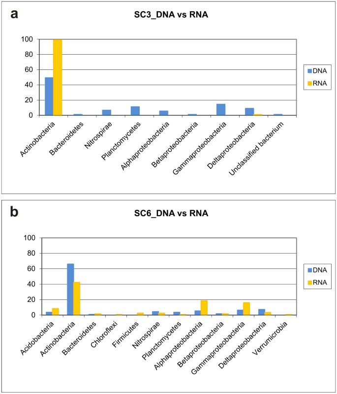
The distribution of different phyla amid the samples is summarized in Figure 4. Only 1 very major phylogenetic group belonging to Actinobacteria was encountered in the clone libraries, which represented 49.4 and 66% of DNA sequences and 99 and 42.iv% of RNA sequences for samples SC3 and SC6, respectively. Besides Actinobacteria, only Planctomycetes and Proteobacteria dominated in both samples, either as full or metabolically agile members. Planctomycetes (11.2%) and Gammaproteobacteria (fourteen.half dozen%) attained some importance in SC3-Deoxyribonucleic acid and Alphaproteobacteria (18.half-dozen%) and Gammaproteobacteria (16.1%) in SC6-RNA.

Distribution of major phylogenetic groups in the 16S rRNA cistron clone library constructed from Tomba della Scimmia SC3 and SC6 samples.
A survey on the sequences obtained from the two samples and the nearest published relatives (Supplementary Tables S1–S4) revealed that 38.half dozen% corresponded to uncultured bacteria from caves and shelters, 26.3% to sequences retrieved from polluted soils and xvi.two% to sequences from different soil types. The data points to the presence of a customs specialized in soil/subterranean niches able to cope with recalcitrant organic affair and pollutants as food sources.
Discussion
The phylum Actinobacteria constitutes a significant part of the bacterial population in subterranean environments8,nine,10. Previous studies on different Spanish and Italian caves revealed that the special microclimatic conditions together with nutrient availability and the nature of the organic matter are important factors controlling the activity of Actinobacteria in cavesix, which take a major role in biogeochemical processes, namely biodeterioration and bioprecipitation of minerals, every bit recently discussed3,4,five,8,9. It is remarkable that 99% of RNA sequences retrieved from sample SC3 were from Nocardia.
The phylum Planctomycetes and the classes Alphaproteobacteria and Gammaproteobacteria, also with some abundance in the samples, are commonly reported in polluted subterranean environmentsxi.
The genus Nocardia is frequently isolated from soils12 and clinical specimens10. In the last few years, members of Nocardia were isolated from European and Asian cavesx. Although the chief reservoir of Nocardia asteroides was thought to be the soil13, Northward. asteroides was common in the air of all the halls and galleries in Castañar de Ibor Cave, Spain, but absent in the air exterior the cavern (unpublished data). Literature data show a worldwide distribution of members of the genus Nocardia in subterranean environments.
The genus Pseudonocardia is also mutual in subterranean environments, such as gold mine cavesx. Pseudonocardia halophobica was institute in Thai caves14. Pseudonocardia spp. were reported in Republic of malta catacombs15, Carlsbad Cavern, New Mexico, The statesxvi and in white colonizations in the Castilian Altamira, Ardales and Santimamiñe caves3,8,17. A previous study on these Spanish caves showed that the DGGE patterns of the metabolically active bacterial communities were equanimous of a major, almost exclusive, band respective to Pseudonocardia, in amounts as high equally 85.6% and 65.7% for Ardales and Santimamiñe caves, respectivelyeight.
The presence and abundance of Nocardia and Pseudonocardia in subterranean environments may be driven past:
- i
Organic matter. Cave colonization by Nocardia and Pseudonocardia were related with agricultural and/or livestock activities on the top soilviii,9,18. The organic carbon content in nearly Italian elevation soils (86.4% of total state surface area) is ≤ 2%xix. Soil organic matter is composed of biodegraded lignin and humic substances, among other macromoleculesxx,21. Subterranean dripping waters contain these macromolecules every bit dissolved organic matter (DOM), which can reach the paintings. Nocardia (east.one thousand. Nocardia erythropolis, Nocardia corallina and Nocardia opaca) and Pseudonocardia spp. use humic substances as sole carbon and nitrogen sources22,23,24. In addition, Nocardia has been shown to dethrone lignin in soil25. Barton et al.16 found that 80% of the total community of a limestone formation in Carlsbad Cavern was represented by Pseudonocardia. These authors suggested that organic matter in this cave is of a phenolic and aromatic nature, equally previously observed in other caves18.
- ii
Phyllosilicates. Phyllosilicates and especially dirt minerals take a loftier adsorbance potential for macromolecules26. DOM from dripping waters may be adsorbed to clay, hindering the loss of food in an oligotrophic environment and in this way may be accessed past leaner. Colonization and growth of Pseudonocardia is favoured by the presence of clayey substrata in the Spanish caves of Ardales and Santimamiñe8. In Carlsbad Cavern, to the limestone that supported the Pseudonocardia were associated dirt particles16. Etruscan paintings were applied on a thin layer of clayii. The stimulation of clays on the colonization and metabolic activeness of Nocardia was well-known fourth dimension ago27.
- iii
Iron oxides. Hematite was used as red pigment for the paintingstwo. Iron is required for an optimal growth of Nocardia 28,29. Hematite and clays tin can adsorbe humic substances, thus facilitating the access of microorganisms to carbon sources30.
In conclusion, the abundant actinobacterial colonization of Tomba della Scimmia is promoted past the presence of organic matter and clays on the walls. Atomic number 26 oxide represents an boosted gene favouring growth. This blazon of colonization was also observed in other Tuscanian tombs with similar constructive features (e.m. Tomba del Colle in the Poggio Renzo Necropolis, Tomba della Pellegrina, near Tomba della Scimmia, etc.). The actinobacteria are involved in the bioprecipitation of minerals4,5,6 (Fig. 3), among other biogeochemical processes, as well as in the biodeterioration of the paintings. In fact, actinobacteria accept a broad ability to produce acids from well-nigh carbohydrates and cause mineral leaching31.
The identification of active microorganisms7,17,32 can provide clues for an effective control and should be the target in cleaning and restoration processes, when possible. Biocides treatments in caves where used in the past and currently are critically discussed because they seem not to be effective or at least have not long-term activity. If we consider the intimate contact between bacteria and mineral layer (Fig. 3), a removal of the dead bacterial biomass, afterwards treatment of the paintings is incommunicable without damaging the pigment layer. This dead biomass will back up the growth of saprophyte fungi as secondary invaders every bit soon as the biocide is degraded or even the biocide used every bit a food source by another bacteria32.
Due to the detail Etruscan pictorial technique (application of pigments on a fine clay layer), the only possible activeness is decision-making the microclimate, visits and the input of DOM to the interior of the tomb past acting on top soil grasses and plants (non agricultural employ, seasonal harvest of spontaneous grasses, etc.) in lodge to reduce organic thing decomposition. Similar deportment were carried out in Altamira Cave with remarkable resultsxviii. Biocide treatments are non recommended in subterranean environments32,33 considering they are not effective at short and mid-term, promote further secondary colonizations and increase microbial biodiversity.
Methods
Sample collection
Sample SC3 and SC6 were nerveless from Tomba della Scimmia on 2nd July, 2009. Both samples represented macroscopic white colonizations located on the paintings (SC3) and on a place without paintings (SC6) (Fig. 2a,b). The samples were taken by scraping off the colonizations with a sterile scalpel. One mL of RNA-later on was added to a set of tubes. Upon collection, the samples were stored on ice and candy or frozen when arrived to the laboratory.
Nucleic acid extraction and PCR amplification
Total nucleic acids from the samples were extracted using the method described by Griffiths et al. 34. Extracted nucleic acids were resuspended in 50 μl of sterile ultrapure water (Sigma-Aldrich). For RNA extraction, a 25 μl aliquot was subjected to a DNase digestion pace using an RNase-Gratuitous DNase (Qiagen) and the RNA was purified using the RNeasy MinElute Cleanup kit (Qiagen), according to the manufacturer's instructions. Complementary Dna (cDNA) from this RNA was synthesized using Superscript Two Opposite Transcriptase (Invitrogen) with a single specific primers: 907R (v′-CCCCGTCAATTCATTTGAGTTT-3′) for bacterial 16S rRNA factor35, Arch 1000R (5′-GGCCATGCACYWCYTCTC-3′) for archaeal 16S rRNA gene36 and ITS4 (v′-TCCTCCGCTTATTGATATGC-3′) for fungal internal transcribed spacer (ITS) regions37, at a temperature of 42°C for 50 min. Distension of cDNA was performed with the bacteria-specific primers, 616F (five′-AGAGTTTGATYMTGGCTCAG-3′)38 and 907R35, the archaea-specific primers, Arch 340F (5′-CCCTAYGGGGYGCASCAG-3′) and Arch 1000R36 and fungi-specific primers, ITS1 (five′-TCCGTAGGTGAACCTGCGG-iii′) and ITS437. One or two μl of cDNA, diluted i/10 were used as a template. PCR conditions were the same for DNA. Amplifications of archaea and fungi from samples SC3 and SC6 were unsuccessful.
PCR reactions were performed in a Bio-Rad iCycler thermal cycler (Bio-Rad). The PCR reaction mixture (ane ml) consisted of 100 μl of PCR buffer (Biotools), xxx μl of MgCltwo (fifty mM, Biotools), ten μl of each primer (fifty mM), 100 μl of dNTPs (100 mM, Bioline), 5 μl of Taq polymerase (Biotools) and 750 μl of sterile ultrapure water. PCR reactions were performed in 0.2 mL PCR tubes containing 25 or 50 μL of reaction mixture and from 0.5 to 2 μl of Dna template (pure or diluted to 2 ng/μl). PCR amplifications were performed using the post-obit thermal conditions: 94°C for 2 min; 35 cycles of 94°C for 15 s, 55°C for xv s (fifty°C for ITS regions), 72°C for 2 min; and a concluding pace of 72°C for 10 min. Positive and negative controls were included in all distension experiments. All amplification products were purified with a JETquick PCR Purification Spin kit (Genomed) and stored at −20°C for further assay.
Cloning and sequencing
DNA libraries of PCR amplified products were constructed using the pGEM-T Easy Vector (Promega) and and then transformed into One Shot Max Efficiency DH5a-T1 chemically competent Escherichia coli (Invitrogen) co-ordinate to manufactureś instructions. Transformants were randomly picked and transferred to multiwell plates containing Luria–Bertani medium supplemented with 100 μg·mL−1 ampicillin and xv% westward/v glycerol and stored at −lxxx°C. We constructed iv libraries. On average 100 clones from each library was sequenced at Macrogen Inc., Seoul, Korea, using the universal bacterial primer 616F.
Sequence analyses
Sequences were checked for bubble by chimera.slayer as implemented in the software package mothur 39. Putative chimeric sequences were excluded from further analysis and 414 sequences were included in phylogenetic analyses. Sequences were aligned using mothur. Afterward this analysis, all sequences were compared to the non-redundant database of sequences deposited at the National Center for Biotechnology (NCBI) and EzTaxon40 using BLASTN algorithm41. Aligned sequences were clustered into operational taxonomic units (OTUs) using mothur, with a 97% sequence identity cutoff. Rarefaction curves were also obtained using mothur. All sequences were submitted to GenBank with consecutive accretion numbers HF58493-HF58701.
Bacterial community fingerprints
Fingerprints of the total and metabolically active microbial communities were obtained by denaturing gradient gel electrophoresis (DGGE) according to Muyzer et al.42. A nested-PCR reaction was performed with the primers 341F (5′-CCTACGGGAGGCAGCAG-3′; with a GC-rich tail added at its 5′-cease) and 518R (5′-ATTACCGCGGCTGCTGG-3′). PCR amplifications were performed using the following thermal conditions: 94°C for 2 min, followed by 35 cycles of 94°C for xv s, 55°C for 15 s and 72°C for thirty s with a terminal extension at 72°C for thirty min. DGGE was carried out using a DCODE™ Organization (Bio-Rad).
Environmental scanning electron microscopy
Textural and microestructural characterization of unlike white colonizations were performed using an Inspect-S50 low-vacuum environmental scanning electron microscope (FEI Company, Nippon) that includes an free energy-dispersive spectroscopy probe. Start the samples were observed and described nether controlled low-vacuum conditions. Farther, the samples were gold sputter covered (gold coater EMITECH K550Y) and observed under high vacuum weather condition to improve the photographic quality and EDS microanalysis.
References
-
Steingraber, S. Abundance of Life: Etruscan Wall Painting (Arsenale, Verona, 2006).
-
Colombini, G. P., Giachi, G., Pallechi, P. & Ribechini, Due east. T. Tecniche pittoriche utilizzate nelle tombe etrusche di Chiusi east Sarteano. In: Pittura Etrusca. Problemi east Prospettive. (ed Minetti A.) 162–167 (Protagon Ed. Toscani Siena, 2003).
-
Cuezva, S., Sanchez-Moral, South., Saiz-Jimenez, C. & Cañaveras, J. C. Microbial communities and associated mineral fabrics in Altamira Cave, Spain. Int. J. Speleol. 38, 83–92 (2009).
-
Cañaveras, J. C. et al. On the origin of fiber calcite crystals in moonmilk deposits. Naturwissenschaften 93, 27–32 (2006).
-
Cuezva, S. et al. The biogeochemical role of Actinobacteria in Altamira Cave, Espana. FEMS Microbiol. Ecol. 81, 281–290 (2012).
-
Piñar, G., Saiz-Jimenez, C., Schabereiter-Gurtner, C., Blanco-Varela, Yard. T., Lubitz, W. & Rölleke, S. Archaeal communities in two disparate deteriorated ancient wall paintings, detection, identification and temporal monitoring by DGGE. FEMS Microbiol. Ecol. 37, 45–54 (2001).
-
Gonzalez, J. M., Portillo, M. C. & Saiz-Jimenez, C. Metabolically active Crenarchaeota in Altamira Cave. Naturwissenschaften 93, 42–45 (2006).
-
Stomeo, F., Portillo, M. C., Gonzalez, J. M., Laiz, L. & Saiz-Jimenez, C. Pseudonocardia in white colonizations in two caves with Paleolithic paintings. Int. Biodeterior. Biodegrad. 62, 483–486 (2008).
-
Groth, I., Vetermann, R., Schuetze, B., Schumann, P. & Saiz-Jimenez, C. Actinomycetes in karstic caves of Northern Spain (Altamira and Tito Bustillo). J. Microbiol. Methods 36, 115–122 (1999).
-
Jurado, Five. et al. Pathogenic and opportunistic microorganisms in caves. Int. J. Speleol. 39, 15–24 (2010).
-
Miller, A. Z. et al. Enigmatic reticulated filaments in subsurface granite. Environ. Microbiol. Rep. 4, 596–603 (2012).
-
Goodfellow, 1000. & Williams, S. T. Ecology of actinomycetes. Ann. Rev. Microbiol. 37,189–216 (1983).
-
Yamamura, H., Hayakawa, Thousand., Nakagawa, Y. & Iimura, Y. Label of Nocardia asteroides isolates from different ecological habitats on the basis of repetitive extragenic palindromic-PCR fingerprinting. Appl. Environ. Microbiol. seventy, 3149–3151 (2004).
-
Nakaew, N., Pathom-aree, Westward. & Lumyong, S. Generic multifariousness of rare actinomycetes from Thai cave soils and their possible use as new bioactive compounds. Actinomycetologica 23, 21–26 (2009).
-
De Leo, F., Iero, A., Zammit, G. & Urzì, C. Chemoorganotrophic bacteria isolated from biodeteriorated surfaces in cavern and catacombs. Int. J. Speleol. 41, 125–136 (2012).
-
Barton, H. A., Taylor, Northward. M., Kreate, M. P., Springer, A. C., Oehrle, S. A. & Bertog, J. L. The bear upon of host rock geochemistry on bacterial customs structure in oligotrophic cave environments. Int. J. Speleol. 36, 93–104 (2007).
-
Portillo, M. C., Saiz-Jimenez, C. & Gonzalez, J. M. Molecular label of total and metabolically active bacterial communities of ''white colonizations'' in the Altamira Cave, Spain. Res. Microbiol. 160, 41–47 (2009).
-
Saiz-Jimenez, C. et al. Paleolithic art in peril: Policy and scientific discipline collide at Altamira Cave. Science 334, 42–43 (2011).
-
Rusco, E., Jones, R. & Bidoglio, Thousand. Organic affair in the soils of Europe: Present status and future trends. Written report EUR 20556.Accessed at http://eusoils.jrc.ec.europa.eu/esdb_archive/eusoils_docs/other/ESF_OM7.pdf on nineteen February 2013.
-
Martin, F., Saiz-Jimenez, C. & Cert, A. Pyrolysis-gas chromatography-mass spectrometry of soil humic fractions. II. The loftier boiling point compounds. Soil Sci. Soc. Am. J. 43, 309–312 (1979).
-
Saiz-Jimenez, C., Hawkins, B. L. & Maciel, Thousand. Due east. Cantankerous polarization, magic-angle spinning 13C nuclear magnetic resonance spectroscopy of soil humic fractions. Org. Geochem. 9, 277–284 (1986).
-
Steinbrenner, G. & Mundstock, I. Untersuchungen zum Huminstoffabbau durch Nokardien. Curvation. Acker. Pflanzenbau Bodenk. 19, 243–255 (1975).
-
Sidorenko, O. D., Aristarkhova, Five. I. & Chernikov, V. A. Changes in the composition and properties of humic acids brought about by the activeness of microorganisms of the genus Nocardia. Biol. Balderdash. Acad. Sci. USSR 5, 150–155 (1978).
-
Solntseva, I. E. Certain energy aspects of microorganism-induced transformation of humic substances. In Humic Substances in the Global Environs and Implications on Man Health (eds Senesi Northward., & Miano T. M.) 373–379 (Elsevier, Amsterdam, 1994).
-
Tuomela, M., Vikman, M., Hatakka, A. & Itävaara, Thousand. Biodegradation of lignin in a compost environment, a review. Bioresour. Technol. 72, 169–183 (2000).
-
Evans, L. T. & Russell, E. West. The adsorption of humic and fulvic acids by clays. J. Soil Sci. 10, 119–132 (1959).
-
Martin, J. P., Filip, Z. & Haider, K. Consequence of montmorillonite and humate on growth and metabolic activity of some actinomycetes. Soil Biol. Biochem. 8, 409–413 (1976).
-
Heim, A. H. & Lechevalier, H. Issue of fe, zinc, manganese and calcium on the growth of diverse strains of Streptomyces. Mycologia 48, 628–636 (1956).
-
Webley, D. M. The consequence of deficiency of iron, zinc and manganese on the growth and morphology of Nocardia opaca. J. Gen. Microbiol. 23, 87–92 (1960).
-
Murphy, E. M., Zachara, J. M. & Smith, S. C. Influence of mineral-spring humic substances on the sorption of hydrophobic organic compounds. Environ. Sci. Technol. 24, 1507–1516 (1990).
-
Abdulla, H. Bioweathering and biotransformation of granitic stone minerals by actinomycetes. Microb. Ecol. 58, 753–761 (2009).
-
Martin-Sanchez, P. M., Nováková, A., Bastian, F., Alabouvette, C. & Saiz-Jimenez, C. Utilize of biocides for the control of fungal outbreaks in subterranean environments: The case of the Lascaux Cave in France. Environ. Sci. Technol. 46, 762–3770 (2012).
-
Saiz-Jimenez, C. Painted Materials. In Cultural Heritage Microbiology (eds Mitchell R., & McNamara C. J.) 3–13 (ASM Press, Washington, DC, 2010).
-
Griffiths, R. I., Whiteley, A. S., O'Donnell, A. Thou. & Bailey, 1000. J. Rapid method for coextraction of Dna and RNA from natural environments for analysis of ribosomal DNA- and rRNA-based microbial community composition. Appl. Environ. Microbiol. 66, 5488–5491 (2000).
-
Teske, A., Wawer, C., Muyzer, G. & Ramsing, North. B. Distribution of sulphate-reducing bacteria in a stratified fjord (Mariager Fjord, Denmark) every bit evaluated by most-probable-number counts and DGGE of PCR-amplified ribosomal Deoxyribonucleic acid fragments. Appl. Environ. Microbiol. 62,1405–1415 (1996).
-
Gantner, S., Andersson, A. F., Alonso-Sáez, Fifty. & Bertilsson, Southward. Novel primers for 16S rRNA-based archaeal customs analyses in environmental samples. J. Microbiol. Methods 84, 12–18 (2011).
-
White, T. J., Bruns, T., Lee, S. & Taylor, J. Amplification and direct sequencing of fungal ribosomal RNA genes for phylogenetics. In PCR Protocols, A Guide to Methods and Applications (eds Innis M. A., Gelfand D. H., Sninsky J. J., & White T. J.) 315–322 (Academic Press, New York, 1990).
-
Zimmermann, J., Gonzalez, J. Grand., Ludwig, W. & Saiz-Jimenez, C. Detection and phylogenetic relationships of a highly various uncultured acidobacterial customs on paleolithic paintings in Altamira Cave using 23S rRNA sequence analyses. Geomicrobiol. J. 22, 379–388 (2005).
-
Schloss, P. C. et al. Introducing mothur: Open source, platform-independent, community-supported software for describing and comparison microbial communities. Appl. Environ. Microbiol. 75, 7537–7541 (2009).
-
Kim, O. South. et al. Introducing EzTaxon-e: a prokaryotic 16S rRNA gene sequence database with phylotypes that stand for uncultured species. Int. J. Syst. Evol. Microbiol. 62, 716–721 (2012).
-
Altschul, S. F., Gish, West., Miller, W., Myers, E. West. & Lipman, D. J. Bones local alignment search tool. J. Mol. Biol. 215, 403–410 (1990).
-
Muyzer, Chiliad., de Waal, Eastward. C. & Uitterlinden, A. G. Profiling of complex microbial populations past denaturing gradient gel electrophoresis analysis of polymerase chain reaction-amplified genes coding for 16S rRNA. Appl. Environ. Microbiol. 59, 695–700 (1993).
Acknowledgements
Thousand.D.-H. was supported by a JAE Enquiry Fellowship from the Spanish Council of Scientific Research and S.C. by a Juan de la Cierva contract. This research was supported with funding provided to the projects CGL2010-17183, 201030E011 and Consolider CSD2007-00058.
Author information
Affiliations
Contributions
C.Due south.-J. conceived, planned the projects and wrote the manuscript. 5.J. collected samples, supervised phylogenetic analysis and performed statistical analyses. M.D.-H. extracted DNA and RNA, performed DGGE and sequence analyses. S.C. and Due south.S.-G. conducted ESEM analyses and interpreted the micrographs. L.L. advised M.D.-H. in laboratory analyses. P.P. and P.T. organized the sampling trips, provided access to the tomb, supplied historical information on the paintings and discussed on the conservation methods.
Ethics declarations
Competing interests
The authors declare no competing financial interests.
Additional information
Accession codes: Sequences were deposited at GenBank nether the accretion numbers HF58493-HF58701.
Electronic supplementary cloth
Rights and permissions
This piece of work is licensed under a Creative Eatables Attribution-NonCommercial-NoDerivs three.0 Unported License. To view a copy of this license, visit http://creativecommons.org/licenses/by-nc-nd/3.0/
Reprints and Permissions
About this article
Cite this commodity
Diaz-Herraiz, K., Jurado, V., Cuezva, Southward. et al. The Actinobacterial Colonization of Etruscan Paintings. Sci Rep three, 1440 (2013). https://doi.org/x.1038/srep01440
-
Received:
-
Accepted:
-
Published:
-
DOI : https://doi.org/10.1038/srep01440
Further reading
Comments
By submitting a comment yous agree to abide by our Terms and Community Guidelines. If you lot find something abusive or that does not comply with our terms or guidelines please flag it every bit inappropriate.
Source: https://www.nature.com/articles/srep01440
0 Response to "Saizjimenez Et Al 2011 Paleolithic Art in Peril Policy and Science Collide at Altamira Cave"
Postar um comentário